Autonomous X-ray Characterization: Pioneering Self-Driving Experiments in Materials Science
Understanding the intricate structures and chemical states of materials is critical to breakthroughs in energy, electronics, and nanotechnology. At our lab, we are advancing the frontier of autonomous experimentation, particularly in X-ray characterization, by developing intelligent systems that can guide and accelerate scientific discovery with minimal human intervention.
Why Automation?
X-ray techniques such as scanning microscopy and X-ray absorption near-edge structure (XANES) spectroscopy are indispensable tools in modern materials research. However, traditional experiments often require exhaustive data collection—leading to time-intensive procedures, sample degradation, and the generation of massive data volumes. Our research focuses on overcoming these limitations by creating AI-driven frameworks that intelligently guide data acquisition, reducing redundancy and enhancing efficiency.
FAST: An Autonomous Workflow for Scanning Microscopy
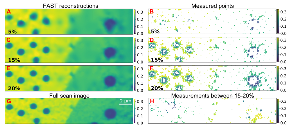
One of our flagship contributions is the Fast Autonomous Scanning Toolkit (FAST), a general-purpose, lightweight, and real-time adaptive scanning framework designed for X-ray and electron microscopy experiments. FAST uses a neural-network-guided dynamic sampling algorithm (SLADS-Net) combined with real-time route optimization and hardware interfacing to direct the beam only to informative regions of the sample. The system continuously updates its internal model and decision-making pipeline based on incoming data, allowing it to identify key features—such as grain boundaries, bubbles, or curvature in 2D materials—without any prior knowledge of the sample.
In a live demonstration at the Advanced Photon Source, FAST successfully imaged WSe₂ thin films with less than 25% of the data points required by conventional methods, all while preserving fine-scale features and enabling curvature analysis. The entire workflow, running on an edge computing device, added less than 2% overhead to experiment time, showcasing the potential for wide deployment at beamlines and beyond.
Smarter Spectroscopy with Bayesian Optimization
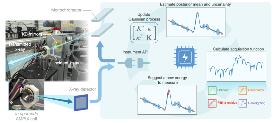
In parallel, we are transforming the way spectroscopic experiments—especially XANES—are performed. Conventional XANES scans often rely on fixed energy grids, which can under-sample key spectral features or over-sample regions with little value. Our approach introduces a domain-informed Bayesian optimization algorithm tailored for XANES, incorporating physical insight about absorption edges, pre-edge peaks, and white lines into the acquisition function itself.
By combining Gaussian processes with engineered acquisition functions sensitive to both statistical uncertainty and known spectral structures, our method achieves remarkable accuracy in locating key spectral features. In tests on real battery and catalyst materials, we reconstructed high-quality spectra with only 15–20% of the typical energy points, achieving sub-0.1 eV accuracy in peak identification—sufficient for tracking dynamic chemical processes in real time.
Real-World Impact and Future Directions
Our work has been successfully deployed at synchrotron beamlines, enabling in-situ and operando studies of battery electrodes and catalysts. The adaptive frameworks not only reduce beam time and measurement load, but also improve time resolution in dynamic studies—critical for studying fast reactions or time-sensitive phenomena.
Looking ahead, we are expanding these methods to:
- Integrate GPU acceleration for ultrafast decision-making,
- Support other X-ray modalities such as fluorescence and transmission,
- Generalize to 3D scanning and multimodal experiments.
By building intelligent, adaptive, and scalable workflows, our lab is helping redefine the boundaries of experimental science—moving toward a future where self-driving laboratories can keep pace with the growing complexity and scale of scientific discovery.
References
[1] Kandel, S., Zhou, T., Babu, A. V., Di, Z., Li, X., Ma, X., Holt, M., Miceli, A., Phatak, C., & Cherukara, M. J. (2023). Demonstration of an AI-driven workflow for autonomous high-resolution scanning microscopy. Nature Communications, 14(1), 5501. https://doi.org/10.1038/s41467-023-40339-1
[2] Du, M., Wolfman, M., Sun, C., Kelly, S. D., & Cherukara, M. J. (2025). Demonstration of an AI-driven workflow for dynamic x-ray spectroscopy. In arXiv [physics.app-ph]. arXiv. http://arxiv.org/abs/2504.17124